
Oral Biopsy
Merheb Surgical Arts IN Peoria & Pekin, IL
The inside of the mouth is typically lined with a special type of skin called mucosa, which is smooth and pink. Any changes in this appearance may indicate a potential pathological process. The following signs could signal the early stages of a pathological condition or cancerous growth:
– Reddish patches (erythroplasia) or white patches (leukoplakia) in the mouth.
– A sore that does not heal and bleeds easily.
– A lump or thickening in the skin lining the inside of the mouth.
These changes can be observed on the lips, cheeks, palate, gums, tongue, face, and neck. Notably, pain is not always present with these conditions, and it is often not associated with oral cancer.
Routine checkups that include oral cancer screenings are essential because early detection is key. Patients with risk factors—such as a history of tobacco and alcohol use, HPV infection, or a family history of oral cancer—should be especially vigilant.
Consultation
During an oral examination, Dr. Merheb can evaluate areas of concern to provide treatment options and take x-rays or a CT scan of the mouth if necessary.
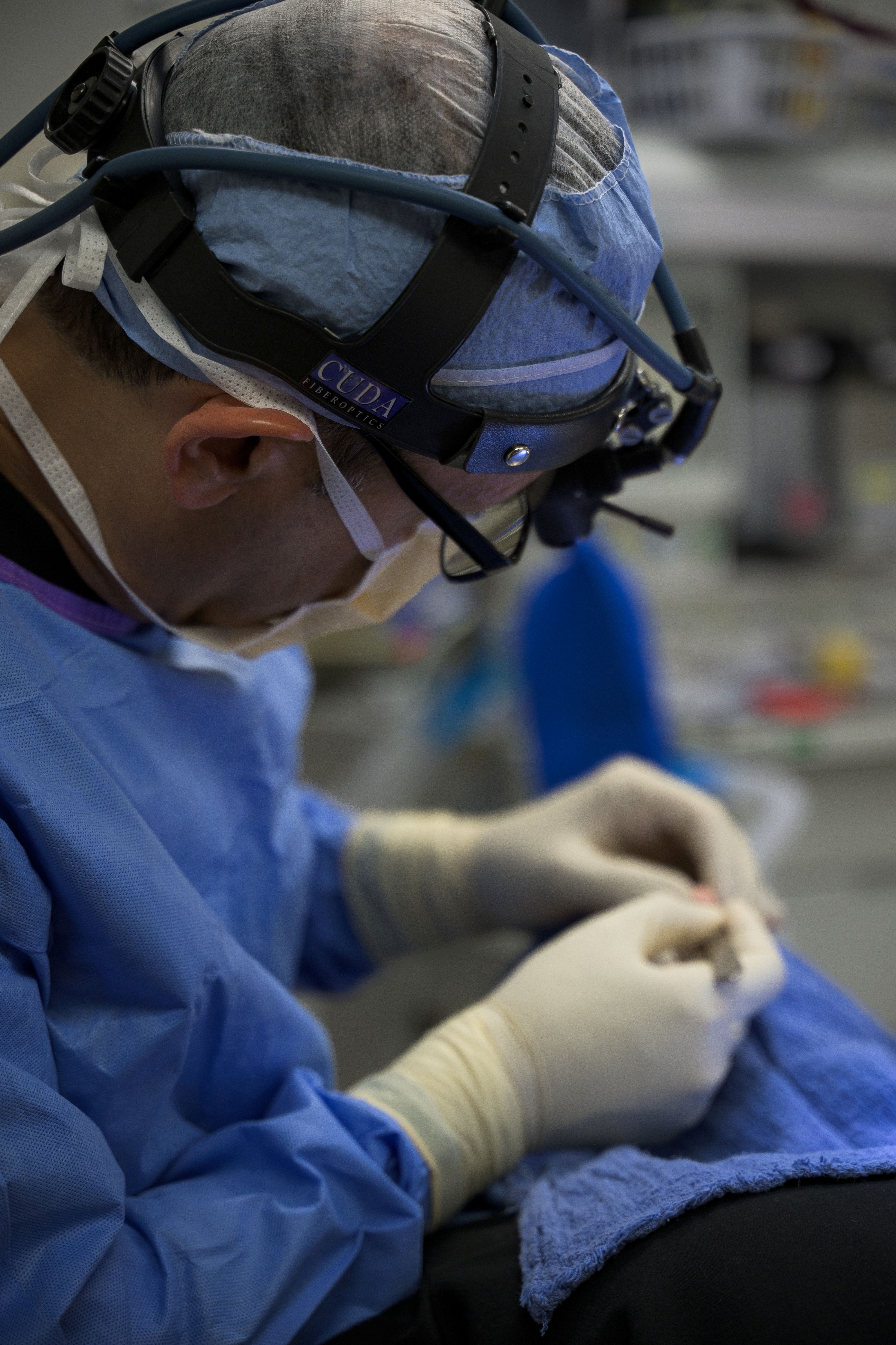
Biopsy
Lesions in the mouth can be managed through various methods, including monitoring and biopsy. A biopsy involves removing a tissue sample from the mouth for examination, typically performed under local anesthesia, and then sending it to your chosen laboratory. Sutures may be applied to control any bleeding. Discomfort is usually minimal, and it is recommended to avoid spicy or acidic foods. Antibiotics and pain medications are often prescribed post-procedure. A follow-up appointment two weeks later allows Dr. Merheb to check on healing and discuss your results. Based on those results, further treatment with Dr. Merheb or other providers may be necessary.